What are the causes of vision loss?
Let's take an intermission prior to discussing Exudative ARMD. Obviously the most important concept of macular disease is vision loss. We all know that good vision was the norm in youth. We all know it becomes a challenge as we age.
Here are the four reasons for vision loss:
- Refractive errors, i.e. need glasses. We use the concept of Best Corrected Vision - BCVA. This is the best vision that can be obtained after having the eye checked for glasses. This obviously is the most common reason for 'vision loss' in young, health individuals. If this is the only problem you have, you do not wind up seeing a retinal specialist.
- Media obstruction, i.e. something is blocking light from entering the eye and getting to the retina where light is transmitted to the brain for interpretation and creation of vision. The most common reason is a cataract or a cloudiness of the normally clear internal lens of the eye. If this is the only cause of vision loss, cataract surgery is the cure. Other reasons for media obstruction are cloudiness of the vitreous cavity (in the back part of the eye) typically due to blood which is a common complication of diabetic retinopathy. The simple rule is that the patient's vision out is equal to the doctors view in of the retina with examination lenses.
- The third is damage to the optic nerve. We're not going to talk about this at this time, however, if there is damage to the optic nerve, the vision loss will be significant.
- The fourth reason for vision loss is what retinal specialists are most focused (sorry) on - damage to the macula must be understood to get an appreciation of the quantity of vision loss, i.e. 20/20 versus 20/40, and the quality of vision loss, i.e. having blind spots and/or distortion.
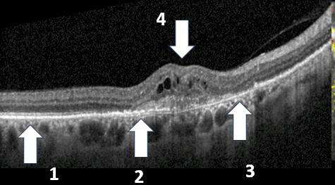
We concentrate on the fovea or the central part of the macula which normally is depressed on the OCT. Here the fovea is swollen from exudation and represents an elevation (4). To assess the vision, we concentrate on the posterior two layers of the retina and RPE. The photoreceptors or cells that transmit light to the retina occupy this space. In the left side of the photo, these layers are relatively normal (1), however in the fovea, the layers are markedly disrupted (2) and even missing (3). GA is present when the RPE is missing (3).
The last concept is that vision loss can be from several reasons - the patient might need glasses, have cataracts, and mild macular damage. Or the cataracts might have been removed and all the vision loss is due to the macular degeneration. I use the concept of macular capability - what would the patient see if the glasses were updated, the cataract removed, and the absence of optic nerve disease. For example if the patient has a 20/100 cataract and I think that the macular capability is 20/40, the patient will not see better than 20/40 after cataract surgery. It's a calculated guess but one based on years of experience. With this assessment, there are no surprises after cataract surgery.