Diagnosis: Optical Coherence Tomography (OCT)
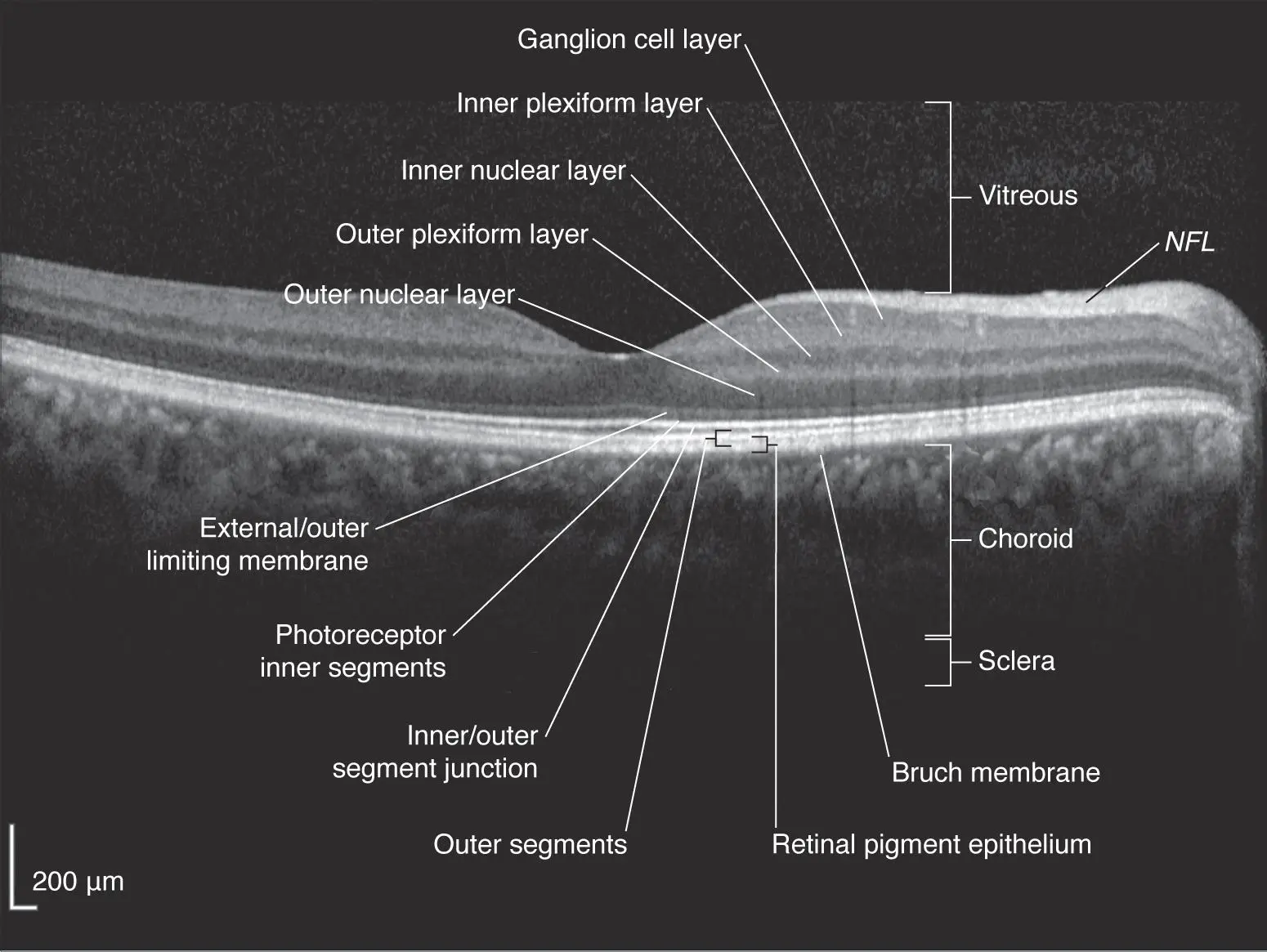
The OCT is one of the most important tests that a retinal ophthalmologist performs in order to diagnose and treat macular degeneration. It is a non-invasive technique that shows a cross-sectional view of the retina. It uses light - it is not an xray. As you can see in this normal OCT, the retina is composed of many layers. In addition to the retina, we also look at the area in front of the retina (vitreous) and behind the retina (choroid).
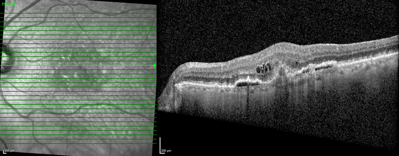
The picture below shows the left macula of a patient with Age-Related Macular Degeneration (ARMD). The photo on the left is how the macula appears when the doctor examines the eye - the optic nerve is on the left edge - the macula is in the central part of the photo. The green line represents the specific area of the macula that is being examined. The corresponding image on the right is the cross sectional view of the retina. One can see irregular thickness and elevations in the center part of the photo. On part 2 of this blog, I will give more information on how to look at an OCT.
The OCT will be performed when you are first examined and then during each subsequent exam to see if the disease has progressed or improved. Ask the photographer or doctor to show your OCT .