Diagnosis: OCT part 2 and OCT Angiography (OCTA)

When we look at an OCT, we look at a cross section of the retina as shown in the left photo. The right photo is the cross section of the macula at the level of the bright green line shown on the left. (1) denotes a relatively normal retina. (2) represents retinal leakage (and thickening) within the retina or intraretinal edema (3) presents neovascularization or abnormal vessels below the retina that are the source of leakage, i.e. wet or exudative macular degeneration (ARMD) and (4) represents a different type of exudative macular degeneration with leakage under the retina. It is not important to understand the different layers of the retina and/or the degrees of leakage but to recognize that the OCT is the most critical test we do in order to diagnose, treat, and follow exudative macular degeneration.
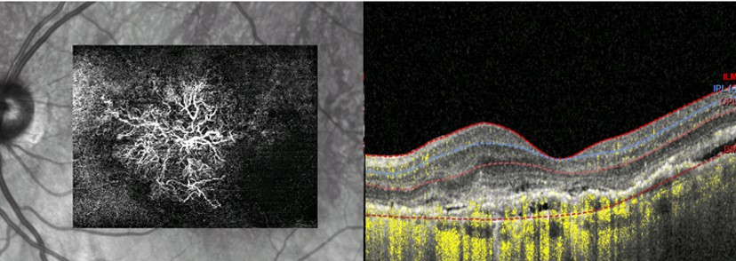
This represents OCT Angiography or OCTA. It is a major advancement on the technology of OCT. Not only can you see the cross section of the macula (on the right) but it actually will show the abnormal neovascularization (vessels responsible for exudation in exudative ARMD) as seen on the left. Not every eye care professional has this device. It is currently not reimbursed by insurance but it adds valuable information in the management of Exudative ARMD especially in patients who have persistent leakage despite multiple injections of anti-VEGF agents.