Diagnosis: Imaging Dyes (IVFA and ICG)
If your doctor finds leakage, or exudation, under your retina and the diagnosis of Exudative Age-Related Macular Degeneration (ARMD) is made, he/she may inject an imaging dye into your vein and take photos of the macula/retina. This test is critical to determine what type of Exudative ARMD you have - there is no 'one size fits all'. There are many phenotypes, or different ways in which the disease presents. This is important because different phenotypes respond to anti-VEGF drugs differently (more later). OCT, OCT Angiography (OCTA), flourescein angiography (IVFA), and indocyanine green (ICG) are all used to best diagnose, treat and follow these diseases.
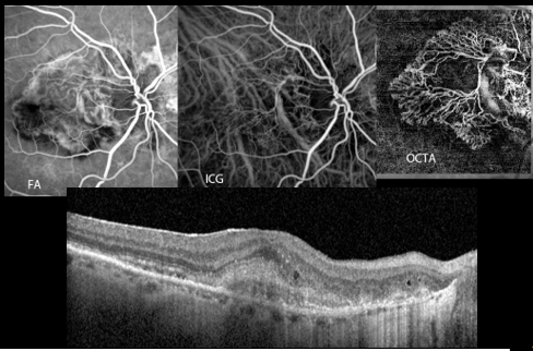
The image above shows how neovascularization can be appreciated on each modality. Below, a cross section of the retina shows neovascularization on OCT. The photo on the top right shows how this neovascularization is appreciated on OCTA, the middle photo by ICG and the top left photo on IVFA.